Topic 4: Transportation Of Minerals Of Living Things – Biology Notes Form Two
The Concept of Transportation of Materials in Living Things
The Concept of Transportation of Materials in Living Things
Explain the concept of transportation of materials in living things
The following is an outline of the importance of transport of materials in living things:
- It facilitates the removal of waste materials from the organism’s body, the excess of whichcould harm an organism.
-
It ensures that essential materials like oxygen, nutrients, water, hormones and mineral saltsare supplied to the cells and tissues as required.
-
It enables essential substances to move from one part of the body to another. For example,food manufactured by photosynthesis in plant leaves is transported from leaves to otherorgans of the plant for use or storage.
Diffusion, Osmosis and Mass- flow
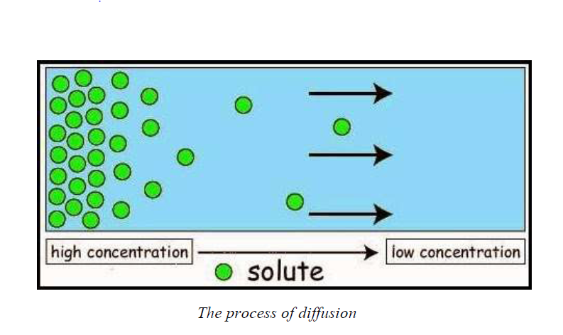
Diagram showing diffusion
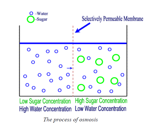
- two solutions with different concentrations; and
- a partially permeable membrane to separate them.
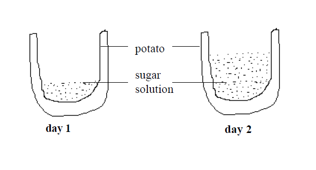
- A little dissolved salt produces a dilute solution with a high water concentration
- A lot of dissolved salt produces a concentrated solution with a low water concentration.
Mass flow
For example, food nutrients from the small intestine have to be moved to cells in the extremities such as toes and fingers, where the nutrient materials have to be transported a long distance.
Materials in higher plants and animals are moved by the process of mass flow. For example, the manufactured food in plant leaves has to be moved to all plant parts, for use or storage, by mass flow.
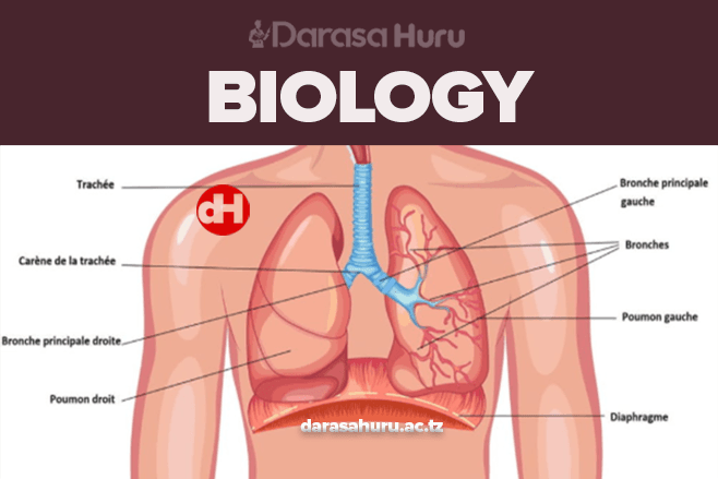
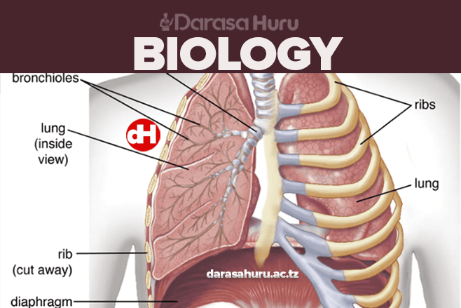
- Gaseous exchange in the lungs of animals and in the leaves of plants
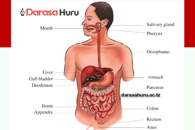
- Absorption of digested food in the ileumThe process of diffusion
- Removal of west materials from cells
- Absorption of nutrients and oxygen into cells
Demonstration of the process of osmosis
The Differences between Diffusion, Osmosis and Mass Flow
Outline the differences between diffusion, osmosis and mass flow
Differences between diffusion and osmosis
Diffusion |
Osmosis |
| It is the movement of all types of substancesfrom the area of their higher concentration tothe area of their lower concentration |
It is the movement of only solvent or waterfrom the area of their higher concentration tothe area of their lower concentration through apartially permeable membrane |
| Diffusion can operate in any medium | Osmosis operates only in a liquid medium |
| Diffusion is applicable to all types ofsubsstances (soilds, liquids and gases) | It is applicable only to solvent part of asolution |
| It does not require any semi-permeablemembrane | A semi-permeable membrane is a must foroperation of osmosis |
| It is purely dependent upon the free energy ofthe diffusing substance | Osmosis is dependent upon the dregree ofreduction of free energy of one solvent overthat of another |
| It helps in equalizing the concentration of thediffusing substance througout the availablespace | It does not equalize the concentration ofsolvent on the two sides of the system |
| Turgor pressure or hydrostatic pressure doesnot normally operate in diffusion | Osmosis is oppossed by turgor or hydrostaticpressure of system |
| It is not influenced by solute potential | Osmosis is dependent upon the solute potential |
| Diffusion of a substance is mostly dependentof the presence of other sustances | It is dependent upon the number of particles ofother substances dissolved in a liquid |
| Factors like water potential, solute potentialand pressure potential do not affect diffusion | Factors like water potential, solute potentialand pressure potential affect osmosis in aliving system |
The Roles of Diffusion, Osmosis and Mass Flow in Movement of Materials in Living Organisms
Explain the roles of diffusion, osmosis and mass flow in movement of materials in living organisms
Through the process of osmosis, nutrients get transported to cells and waste materials getmoved out of them.
Transport of Materials in Mammals, the Structure of the Mammalian Heart
The external structure of the mammalian heart is as shown in the labelled diagram below:

The walls of the ventricles are thicker than those of the auricles because
The functions of each part and the associated structures are as follows:
INTERNAL STRUCTURE OF THE MAMMALIAN HEART
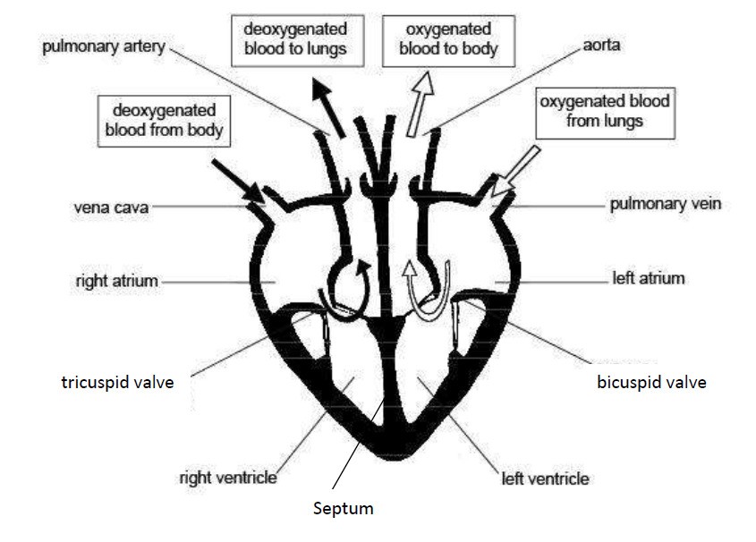
The heart has several valves. And these valves have flaps that ensure that blood flows in one direction only.
These valves include the following:
- The tricuspid valve; found between the right auricle and right ventricle
- The bicuspid valve: found between left auricle and left ventricle
-
Semi-lunarvalves which are located at the bases of the pulmonary artery and aortato preventblood from flowing back into the ventricles.
Functions of parts of the mammalian heart
Part of the heart |
Function |
| Aorta | The largest artery in the body; it conducts freshly oxygenated bloodfrom the heart to the tissues. |
| Superior vena cava | Large vein that brings deoxygenated blood from the upper parts ofthe body to the right atrium |
| Inferior vena cava | Large vein that brings deoxygenated blood from lower regions of thebody to right atrium |
| Pulmonary artery | Carries deoxygenated blood from the right ventricle to the lungs |
| Pulmonary vein | Blood vessel that carries oxygenated blood from the lungs to the leftatrium |
| Right atrium | This chamber of the heart receives deoxygenated blood from thebody (from the superior and inferior vena cava). |
| Left atrium | This chamber of the heart receives oxygenated blood from the lungs |
| Tricuspid valve | Located on the right side of the heart between the right atrium (RA)and right ventricle (RV) |
| Bicuspid valve | Located on the left side of the heart between the left atrium (LA) andthe left ventricle (LV) |
| Right ventricle | The chamber of the heart that pumps deoxygenated blood to thelungs |
| Left ventricle | Receives blood from the left atrium and pumps it into the aorta fortransport to the body cells |
| Septum | Divides the right and left chambers of the heart |
The Adaptations of the Parts of the Mammalian Heart to their Functions
Explain the adaptations of the parts of the mammalian heart to their functions
The heart is adapted to carry out its functions by having the following features:
The cardiac muscle is adapted to be highly resistant to fatigue.
The presence of the cardiac muscles enables the heart to beat rhythmically.
The pericardium which surrounds and protects the heart from physical damage.
Pericardial fluid which prevents friction when the heart beats.
Bicuspid and tricuspid valves between atria and ventricles which prevent the backflow ofblood.
Septum which prevents the mixing of deoxygenated blood in the right and oxygenated bloodin the left chambers of the heart.
Its own blood supply for supplying nutrients and removing waste.
The left ventricle has thick muscular wall to pump blood at a higher pressure to the distantbody tissues,
The heart is supplied with the nerves which control the rate of heartbeat depending on thebody requirements.
Blood vessels
The Structure of Arteries, Veins and Capillaries
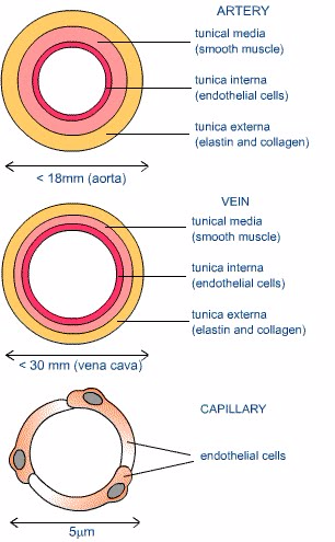
Describe the structure of arteries, veins and capillaries
- Capillaries consist of anendothelium whichis only one cell thick.
- Walls of arteries and veins consist of 3 layers.
- The inner layer consists of a thin layer of endothelial cells.
-
Themiddle layer is made up of smooth muscle with some elastic fibres. Thislayer controls the diameter of the vessel and hence the amount of bloodand its rate of flow.
- The outer layer is composed of connective tissue; this holds the blood vessels in place in the body.
Detailed structure
Arteries
The walls of arteries are much thicker as it carries blood away from the heart at high pressure.
Arteries are near the surface of the skin; the changes in the arteries diameter can be felt as a pulse.
The walls have fewer elastic fibres and the lumen is wider (to allow for easier blood flow).
The one cell thick endothelial layer is a continuation of the lumen arteries and veins.
Diffusion is a relatively slow process and hence the structure of capillaries is suited to slowing down the flow of blood.
They form an expansive blood flow network, such that no cells are far from blood supply
How different blood vessels are adapted for their function
Blood vessel |
Function |
Adaptation |
| Artery | Carries blood away from heart at high pressure | Thick, elastic, muscular walls to withstand pressure and to exert force (pulse) |
| Vein | Returns low-pressure blood to heart | Large diameter to offer least flow resistance. Valves to prevent back flow. |
| Capillary | Allows exchange of materials between blood and tissues | Thin, permeable walls |
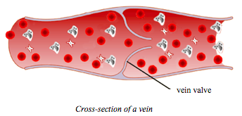
Cross section of vein
Differences between arteries, veins and capillaries
Arteries |
Veins |
Capillaries |
| All arteries carry bloodawayfrom the heart | All veins carry bloodtowardsthe heart | Capillaries carry bloodfrom arteries to the veins |
| With the exception of the pulmonary artery, all arteries carryoxygenatedblood | With the exception of the pulmonary vein, all veins carrydeoxygenatedblood | Bloodslowly loses its oxygen |
| They carry blood which is usuallyrich in digested foodmaterials | Except for the hepatic portal vein, they carry blood which usuallyhaslittledigested foodmaterials | Bloodslowly loses its food |
| Have relatively narrower lumens (see diagrams above) | Have relatively wide lumens (see diagrams above) | Have relatively narrow lumens (see diagrams above) |
| Have relatively athicklayer of muscles and elastic fibres | Have relatively athinlayer of muscles and elastic fibres | Theydo not havemuscles and elastic fibres |
| They havethick outer walls | They havethin outer walls | Walls are onlyone cell thick |
| They carry blood athigh pressure | They carry blood atlow pressure | Pressure gradually fallsas blood flows from arteries to veins |
| Do not have valves (except for the semi-lunar valves of the pulmonary artery and the aorta) | Have valves throughout the main veins of the body to prevent the back flow of blood. | Have no valves |
| Have bright red blood (because it is rich in oxygen) | Brown-red blood | Brown-red blood |
| Located deep in the to body surface | Located near to body surface | Capillaries are found inside all tissues |
| Walls arenot permeable | Walls arenot permeable | Walls arepermeable |
| Blood flows in pulses | Nopulse | Pulse gradually disappears |
Simple Experiments to Determine Pulse Rates in Human Being
Carry out simple experiments to determine pulse rates in human being
Activity 1
Carry out simple experiments to determine pulse rates in human being
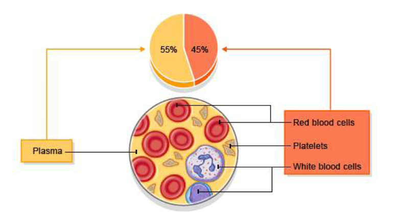
The Blood
- Plasma which is a clear extracellular fluid.
- The solid component, which are made up of the blood cells and platelets
- Erythrocytes, also known as red blood cells (RBCs)
- Leukocytes, also known as white blood cells (WBCs)
- Platelets, also known as thrombocytes
Blood cells

The Function of Major Blood Components
Explain the function of major blood components
Red blood cells (RBCs) have two main functions:
- To pick up oxygen from the lungs and deliver it to tissues elsewhere.
- To pick up carbon dioxide from other tissues and unload it in the lungs.
- Phagocytes: Engulf and digest invading bacteria and viruses (pathogens). It is the body’smain defence against germs (microbes).
-
Lymphocytes:produce antibodies which neutralize antigens from bacteria or viruses.Theykill microbes or make them clump together, to be removed in thelymph glands.
White blood cells are produced in the yellow marrow of the bone, spleen, thymus and lymphaticsystem.
- Secrete vasoconstrictors which constrict blood vessels, causing vascular spasms in brokenblood vessels.
- Form temporary platelet plugs to stop bleeding.
- Secrete procoagulants (clotting factors) to promote blood clotting.
- Dissolve blood clots when they are no longer needed.
- Digest and destroy bacteria.
- Secrete chemicals that attract neutrophils and monocytes to sites of inflammation.
- Secrete growth factors to maintain the linings of blood vessels.
- Plasma serves as a transport medium for delivering nutrients to the cells of the various organsof the body.
- It transports waste products derived from cellular metabolism to the kidneys, liver, and lungsfor excretion.
- It fights infections since it contains antibodies.
- It is also a transport system for blood cells, and it plays a critical role in maintaining normalblood pressure.
-
Plasmahelps to distribute heat throughout the body and to maintainhomeostasis, orbiological stability, including acid-base balance in theblood and body.6. It carries and transports some hormones.
The Effects of HIV on White Blood Cells
Explain the effects of HIV on white blood cells
Blood Groups and Blood Transfusion
Serum is blood plasma without fibrinogen. It can be stored without clotting, and is used intransfusions.
Blood group |
Antigen |
Antibodies |
Agglutinates |
| A | A | Anti-B | Anti-A serum |
| B | B | Anti-A | Anti-B serum |
| AB | A and B | None | Anti-A and anti-B serums |
| O | None | Anti-A and anti-B | Neither serum |
Blood grouping
The Relationship between Blood Groups and Blood Transfusion
Outline the relationship between blood groups and blood transfusion
Recipient |
Donor |
|||
| A | B | AB | O | |
| A | √ | × | × | √ |
| B | × | √ | × | √ |
| AB | √ | √ | √ | √ |
| O | × | × | × | √ |
Note: a tick (√) means compatible and a cross (×) means incompatible.
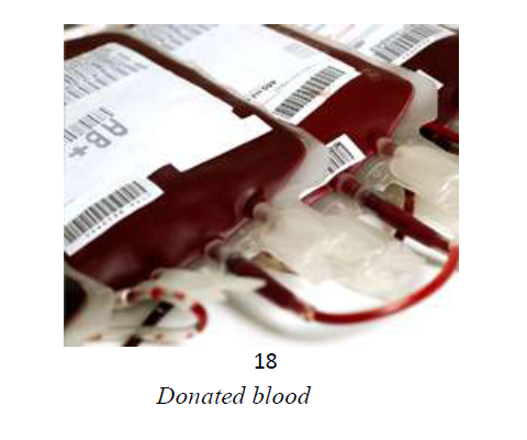
Blood transfusion has a number of advantages. These are some of the benefits the donated bloodcan provide for patients in need:
Provide red cells and platelets for patients with blood disorders such as sickle cell.
Disadvantages of blood transfusions
Medical reactions:
The precautions may include the following:
During blood transfusion, vital signs such as body temperature, heart rate, and bloodpressure are carefully monitored.
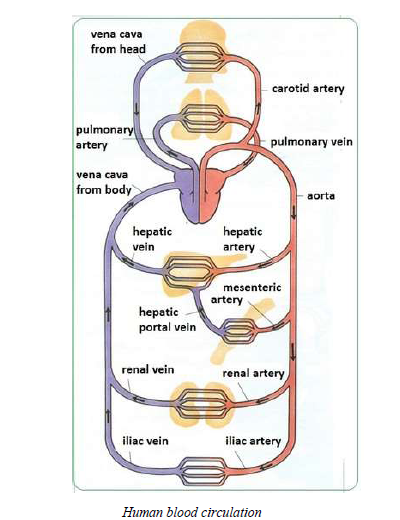
Blood Circulation
Blood Circulation in Humans
Describe blood circulation in humans
The Importance of Blood Circulation in Humans
Explain the importance of blood circulation in humans
-
Everycell in the body needs to received oxygen and nutrients. Blood rich inoxygen is sent tothe body organs, tissues and cells to nourish themthrough blood circulation.
- It enables transportation of waste products from body tissues to excretory organs so as to beremoved from the body.
- Protects the body against diseases and infections through the white blood cells.
- Facilitates blood clotting to prevent loss of blood.
- Maintains body temperature by distributing body heat evenly from the liver and spleen to allbody parts.
Disorders and Diseases of the Human Blood Circulatory System
Mention disorders and diseases of the human blood circulatory system
Additional notes on diseases and disorders of the circulatory system:
Causes and effects of diseases and disorders of the human vascular system
Disease / Disorder |
Description |
Causes |
Effects / Symptoms |
|
| 1 | Anaemia | A reduction inthe quantity of(oxygencarrying)haemoglobin inthe blood and/orbelow normalquantity of redblood cells. | The many possible causesinclude:1.Haemorrhagicanaemia – due to lossof blood2.Iron-deficiencyanaemia – due toinsufficient iron, oftendue tdietarydeficiency.3.Haemolytic anaemiaresult from theincreased destructionof red blood cells e.g.due to toxic chemicals,autoimmunity, theaction of parasites,abnormal forms ofhaemoglobin orabnormal red bloodcells.4.Anaemia can also becaused by the impairedproduction of red bloodcells, as in leukaemia(when red blood cellproduction in the bonemarrow is suppressed). |
Main symptoms are:Excessivetiredness Breathlessnesson exertion Pallor (i.e.looking pale, esp.on face andpalms) Low resistance toinfe |
| 2 | Angina | Pain afterphysical effort | Narrowed coronary arteriesbeing unable to supplyincreased blood flow requiredfor increased physicalexertion. (The arteries mayhave been narrowed by theaccumulation of atheromatousplaque – see atherosclerosis,below.) |
Typical symptomsinclude short-termdiscomfort such as anache, pain or tightnessacross the front of thechest when orimmediately followingexertion or othersituations in which heartrate is increased e.g. dueto panic or an argument.Other less commoneffects & symptoms arealso possible e.g. similarpain when or soon aftereating. |
| Aneurysm | Balloon-likebulge or swellingin the wall of anartery | In general, causes can begenetic or due to disease, e.g.1. a degenerative disease a syphilitic infection -causing damage to themuscular coat of theblood vessel2.a congenital deficiencyin the muscular wall |
Aneurysms can causethe wall of the bloodvessel to weaken. Whenan aneurysm gets biggerthe risk of ruptureincreases. That can leadto severe haemorrhage(bleeding) and othercomplications – some ofwhich may be lifethreatening. |
|
| 3 | Arteriosclerosis | Hardening of thearteries.(Arteriolosclerosis is thehardening ofarterioles.)Artery wallsthicken, stiffenand loseelasticity, aprogressivecondition thattypicallyworsens overtime unlessaction is taken toaddress it.Note: Healthyarteries areflexible andelastic. |
High blood pressure (alsoknown as hypertension) iswidely cited as a cause of, orat least a contributory factorto, the development ofarteriosclerosis.To reduce risk, keep bloodpressure within a healthyrange. See also how to reducerisk of atherosclerosis (below). |
Arteriosclerosis (incombination withatherosclerosis orotherwise) can reducethe flow of blood,hence the supply ofoxygen, nutrients etc.,to tissues in theaffected area.Arteriosclerosis canaffect any artery in thebody but is of greatestconcern when occurs inthe heart (coronaryarteries) or the brain. |
| 4 | Atherosclerosis (Atheroma)- a commontype ofarteriosclerosis (see above) | •Multiple fatty plaques(consisting of e.g.cholesterol andtriglyceride)accumulate on theinner walls of arteries.To reduce risk:1. Eat sensibly (seebalanced diet)2.Don’t smoke3. Take appropriateregular exercise4. Maintain a healthybody weight5. Do not consumeexcessive alcohol |
A chronic disease thatcan remainasymptomatic fordecades. However,blood flow is restrictedand eventuallyobstructed.Various complicationsof advancedatherosclerosis arepossible. One of themost significant risks isof an infarction due tosoft plaque suddenlyrupturing, causing theformation of a thrombus(blood clot) that canslow or stop blood flowleadingto death of thetissues fed by the artery.Thrombosis of acoronary artery cancause a heart attack(Myocardial infarction).The same process in anartery to the brain iscommonly calledstroke.6. CoronarythrombosisA thrombus is ablood clot.Thrombosis is acondition inwhich bloodchanges from aliquid into aCoronary thrombosis canoccur due to the accumulationof fatty deposits (plaques)inside the arteries, i.e.atherosclerosis. The hardeningof arteries (arteriosclerosis)can also contribute to reducedCan lead to a |
|
| 6. | Coronarythrombosis | A thrombus is ablood clot.Thrombosis is acondition inwhich bloodchanges from aliquid into asolid state,producing a ‘clot'(thrombus).Coronarythrombosis isthe condition inwhich thethrombus isformed in one ofthe 3 majorcoronary arteriesthat supply theheart. |
Coronary thrombosis canoccur due to the accumulationof fatty deposits (plaques)inside the arteries, i.e.atherosclerosis. The hardeningof arteries (arteriosclerosis)can also contribute to reducedblood flow leading to coronarythrombosis. |
Can lead to amyocardial infarction(heart attack).Sensations that might beindications of coronarythrombosis leading tomyocardial infarctioninclude: sudden sharppain behind thesternum (breastbone) sudden sharppain on the lefthandside of thechest, that mightspread down theleft arm pain radiatingtowards the jaw,ear, handsstomach, rightarm constrictingsensation in thethroat areadifficultybreathing sudden, severedizziness and/orfainting,experienced withpain. |
| 7. | Haemophilia | Blood clots onlyvery slowly. | Deficiency of either oftwo blood coagulationfactors:o Factor VIII(antihaemophilic factor), oro Factor IX(Christmasfactor) Hereditary -symptoms in males;may be ‘carried’ byfemales who can pass itto their sons withoutbeing affectedthemselves. |
The person mightexperience prolongedbleeding after any injurythat causes an openwound. In severe casesof haemophilia theremay be spontaneousbleeding into musclesand joints.Treatment: Bleeding incases of haemophilia hasbeen treated bytransfusions of plasmacontaining the missingfactor, or withconcentratedpreparations of FactorVIII or Factor IXobtained by freezingfresh plasma. |
| 8 | Haematoma | A collection oraccumulation ofblood outside theblood vessels,which may clotforming aswelling. | The different types ofhaematoma generally havedifferent causes: An intracerebralhaematoma may bedue to a head injury. A perinealhaematoma may occurdue to bleeding from avaginal tear orepisiotomy (cut) duringchildbirth. |
Effects & symptomsalso depend on the typeof haematoma: An intracranialhaematomamight compressthe brain andincrease pressurewithin the skull A subduralhaematoma canbe lifethreatening |
| 9.Haemorrhoids | Haemorrhoids(also called’piles’) areswellingscontainingenlarged andswollen bloodvessels in oraround therectum and anus. | Risk factors – rather than directcauses – include: excessive body weight prolonged constipatione.g. due to insufficientdietary fibre. prolonged diarrhoea lifting heavy objectsfrequently pregnancy – which canplace increasedpressure on pelvicblood vessels, thoughhaemorrhoids oftendisappear after thebirth age (above 50 years) family history ofhaemorrhoids (geneticpredisposition) |
Symptoms ofhaemorrhoids caninclude: Bleeding (brightred blood) afterpassing a stool A pile movingdown, outside ofthe anus(prolapse) a mucusdischarge afterpassing a stool itchiness aroundthe anus soreness andinflammationaround the anussensation ofbowels still beingfull and in needof emptying |
Practical Exercises to Measure Human Pulse Rate and Blood Pressure
Carry out practical exercises to measure human pulse rate and blood pressure
Activity 2
Carry out practical exercises to measure human pulse rate and blood pressure
The Lymphatics System
The Concept of Lymphatics
Explain the concept of lymphatics
The Human Lymphatic System
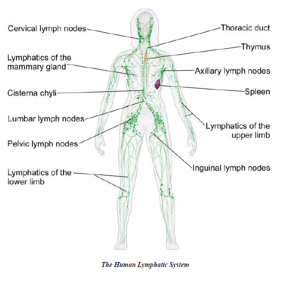
Functions of the lymphatic system
The lymphatic system performs the following functions:
- Removes excess fluid and waste products from the interstitial spaces between the cells andreturns it into the bloodstream.
-
Italso functions in transporting white blood cells to and from the lymphnodes into thebones, and antigen-presenting cells (APCs), such asdendritic cells, to the lymph nodes wherean immune response isstimulated.
-
Special lymph vessels (lacteals) absorb fat andfat-soluble vitamins from the small intestineand deliver these nutrientsto the cells of the body where they are used by the cells.
-
Protectsthe body against germs. Lymph glands produce lymphocytes whichproduceantibodies that fight against microbes. They also containphagocytes, which eat dead whitecells and microbes in the lymph.
The Common Disorders and Diseases of the Lymphatic System
Mention the common disorders and diseases of the lymphatic system
-
Filarialinfection can cause lymphoedema of the limbs, genital disease(hydrocele, chylocele,and swelling of the scrotum and penis). It alsocauses recurrent acute attacks, which areextremely painful and areaccompanied by fever.
- The infected people may have lymphatic and kidney damages.
- Sometimes the swollen limbs become infected.
- The infected person is disabled and cannot work to earn his/her living.
Causes, Symptoms, Effects and Prevention of Disorders and Diseases of the Human Lymphatic System
Explain causes, symptoms, effects and prevention of disorders and diseases of the human lymphatic system
Effective treatment and preventive efforts would include:
- spraying insecticides to kill mosquitoes;
- giving antibiotics to prevent or control infection;
- giving medications to kill microfilariae circulating in the blood;
- applying pressure bandages to reduce swelling; and
- surgically removing infected tissue.
The Concept of Vascular System
Explain the concept of vascular system
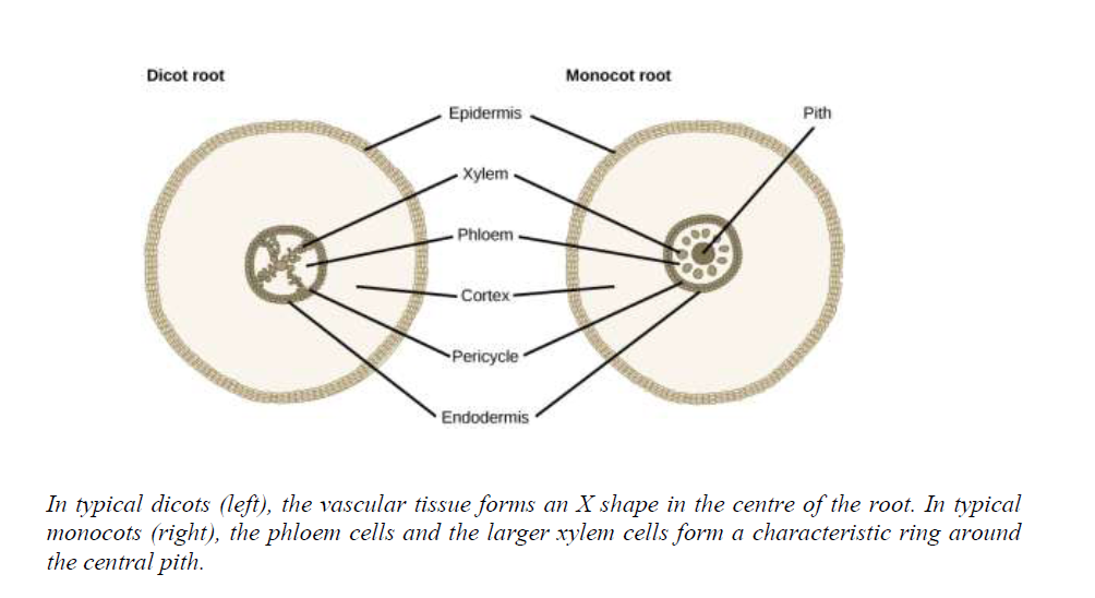
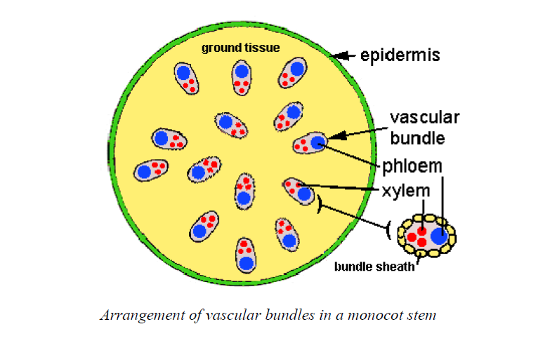
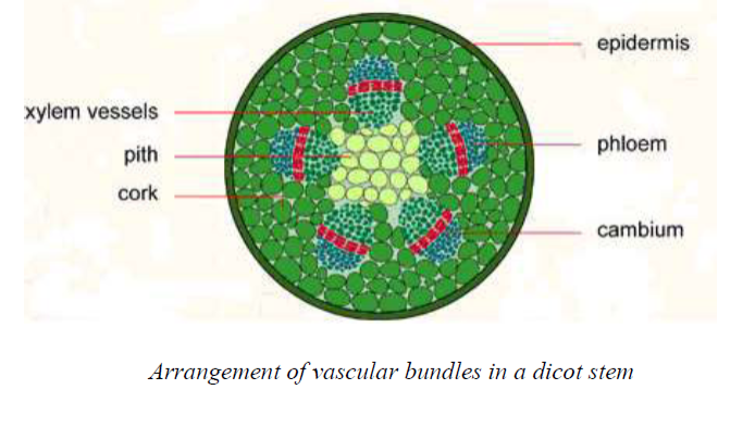
Components of Vascular System
Describe components of vascular system
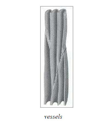
Vessels
The end walls of the vessels are eitherpartially or fully dissolved.
Phloem
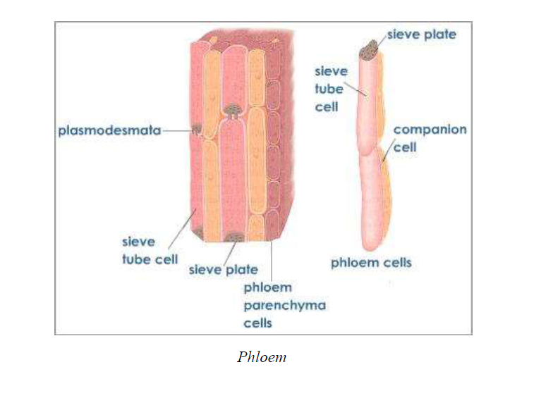
Sieve tubes
- Provides support for woody plants.
- Transports water and solutes from roots to all plant parts.
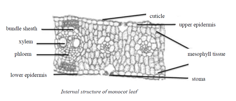
Absorption and Movement of Water and Mineral Salts in Plants
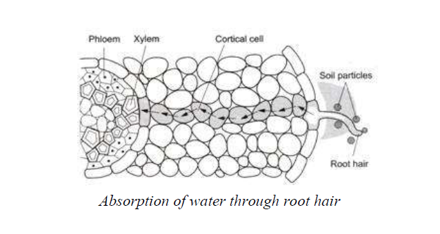
Root pressure
Cohesion and adhesion forces maintain a continuous column of water in the xylem vessels fromthe roots to the leaves of plants.
Cooling of the plant
Maintenance of turgidity
Transpiration is a necessary evil because of the following facts:-
- A large amount of absorbed water is lost during transpiration which is harmful to plants.
- Unnecessary wastage of energy takes place during the process of water absorption which islost due to transpiration.
-
Whenthe rate of transpiration is high in plants growing in soil deficientin water, an internalwater deficit develops in plants which may affectmetabolic process.
- Many xerophytes undergo structural modifications and adaptations to check transpiration.
Size of leaves
Epidermal hairs
Environmental factors
Temperature
Wind
Soil water