TOPIC 3: COORDINATION - BIOLOGY NOTES FORM THREE
COORDINATION
Coordination is the working together of different parts of the body in an orderly and organized manner
Without coordination the body becomes disorderly and it may fail to function properly.
IMPORTANCE OF COORDINATION
1. Coordination ensures survival of organisms.
2. Coordination enables organism to detect their life necessities such as food for heterotrophs, detection of light by autotrophs.
3. Coordination helps living organism to respond to their stimuli.
IRRITABILITY OR SENSITIVITY
Is the ability to perceive, interpret and respond to changes in the internal and external environment.
1. External environment
This is outside surrounding of whole organisms.
Components of external environment
The following are components of external environment:
i. Light
ii. Sound
iii. Pressure
iv. Gravity
v. Chemicals
vi. Water
vii. Food
2. Internal environment
This is the surrounding at cells within the body of an organism.
Components of internal environment
The following are components of internal environment:
i. Water
ii. Glucose
iii. Minerals
iv. Ions
v. pH
vi. Temperature.
COMPONENTS OF COORDINATION
There are five components of coordination namely:
i. Stimulus
ii. Receptors
iii. Coordinators
iv. Effectors
v. Response
I. STIMULUS ( plural: Stimuli)
Is the change in the environment of an organism
TYPES OF STIMULI
There are two types of stimuli, namely:-
a. External stimulus
b. Internal stimulus
A. EXTERNAL STIMULUS
Is the stimulus which is associated with the surrounding environment
Example of external stimuli
> Heat
> Wind
> Pressure
> Chemicals
> Water
> Food
> Light
B. INTERNAL STIMULUS
Is the stimulus which occurs within the organism
Example of internal stimuli
> Water
> Glucose
> Mineral ions
> pH
> Temperature
II. RECEPTORS
Are the specialized cells that detect stimulus
In animals receptors are located in specialized organs known as sense organs.
Example of receptors
> Receptor for pain, touch, heat, and cold-are located in the skin
> Receptors for taste-located in the tongue
> Receptors for light-located in the eye
> Receptors for sound-located in the ear
> Receptors for smell-located in the nose
— When a receptor detects stimuli, it creates impulses which are transmitted to the coordinating system through nerve cells.
NERVE IMPULSE
Is a slight electric charge which travels along a nerve cell
III. A COORDINATOR
Is an organ that receives messages from the receptors, translates them and sends the information back to effectors for action.
Example of coordinators
1. The brain
2. Spinal cord
IV. EFFECTORS
Are the parts of the body that respond to the stimuli.
Example of Effectors
1. Muscles
2. Glands
3. Cilia
4. Flagella
V. RESPONSE
Is a behavioural, physiological or muscular activity initiated by a stimulus
OR
Is the change shown by an organism in reaction to a stimulus
Examples of response
— Blinking when an insect lands on the eye
— Dropping a hot object.
The table below shows the relationship between some stimuli, receptor, effectors and response
| Stimuli | Receptors | Effectors | Responses |
| Heat | Skin | Skin | Secretion of sweat, sweating |
| Cold | Skin | Skeletal muscles | Uncontrolled contraction and relaxation of skeletal muscles, shivering |
| Skin | Formation of goose pimples. | ||
| Taste | Tongue | Salivary glands | Secretion of saliva, salivation |
| Pain | Skin | Skeletal muscles | Contract, move organs away from source of pain |
| Sound | Ear | Ear drum | Hearing of noise, music or sound. |
The sequential order of transmissions of a nerve impulse from a sensory organ to the organism’s response is
![]()
The Ways in Which Coordination is Brought About
Coordination is controlled or effected by two major systems, namely:
i. Nervous system
ii. Hormonal system called endocrine system
> The coordination in simple multi-cellular animals is controlled by nervous system only.
> The coordination in higher animals called vertebrates (including human beings) is controlled by nervous system and endocrine system.
> Coordination in plants is under the control of hormones.
NERVOUS COORDINATION IN HUMAN, NEURONES
NEURONES: Are cells which carry electrical impulses from the central nervous system to all parts of the body
Neurone is the basic unit of the nervous system.
Neurones are also called nerve cells
STRUCTURE AND FUNCTIONS OF THE NEURONE
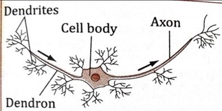
Each neurone consists three basic features, namely;-
i. The cell body
ii. Dendrites
iii. The axon
Other parts/features of the neurone are:
(i) Dendrons
(ii) Myelin sheath
(iii) Schwann cells
(iv) Node of Ranvier
(v) Axoplasm

CELL BODY
Is the main part of the nerve cell
Function of the cell body
i. It gives rise to other parts of the nerve cell
ii. It is the main control centre of the nerve cell
Components of the cell body
The cell body has the following components:
(i) Cytoplasm– enclosing the nucleus.
(ii) Nucleus– which control all activities
(iii) Mitochondria– that provide energy for metabolic processes.
DENDRITES
Are short numerous fibres which receive nerve impulses from other neurones and transmit them to the cell body.
THE AXON
Is the elongated fibre that extends from the cell body The longer the axon, the faster it transmits information.
The role of axon
It transmits nerve impulses away from the cell body.
MYELIN SHEATH
Is a fatty layer that covers axon for protection and insulation
Function of myelin sheath
i. Protects the neuron and allow impulses to travel faster.
ii. It insulates the axon
SCHWANN CELLS
Are cells found on the surface of myelin sheath
Function of Schwann cells
i. They secrete the myelin sheath
NODES OF RANVIER
Are constrictions which interrupt myelin sheath at exactly one millimeter interval
Role/function of node of ranvier
i. Used to speed up the transmission of impulses.
DENDRONS
Are extensions of the cell body
They form branches known as dendrites
Function of Dendrons
i. They transmit impulses towards the cell body
AXOPLASM
Is a specialized type of cytoplasm which is continuous with the cytoplasm in the cell body
Function of axoplasm
i. It is a part through which nerve impulses travels
NEURILEMMA
Is a layer of cells which encloses the myelin sheath
ADAPTATION OF NEURONES TO THEIR FUNCTION
1. They have numerous mitochondria for energy supply during conduction of impulses
2. They are long so as to enables transmission of impulses to long distance in the body.
3. They have node of Ranvier to increase the speed of impulse transmission
4. They are supplied with denser network of blood capillaries for supply of food and oxygen
5. They are numerous for effective transmission of impulses along the whole body.
6. They are covered with fatty myelin sheath for protection and insulation.
7. They have numerous dendrites for connectivity with other neurons.
8. They have Schwann cells that secrete myelin sheath.
9. They have elongated axons which help in quick transmission of impulses.
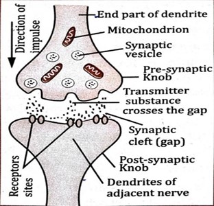
NB: The axon terminates into synaptic knobs.
These knobs have vesicles containing a chemical transmitter substance, for example acetylcholine.
The axon of one neuron and the dendrites of the next neuron do not actually touch each other.
The gap between neurons is called the synapse
A SYNAPSE
Is a junction between two neurones
Function of synapse
i. It enables impulse to be passed from one neurone to another.
ii. It ensures that impulses are transmitted in one direction only.
THE TRANSMISSION OF NERVOUS IMPULSES ACROSS A SYNAPSE
The transmission of nervous impulses across a synapse is mediated by chemical substances called neurotransmitter
Example of neurotransmitters
i. Acetylcholine
ii. Noradrenaline
The transmission of nervous impulses across synapses occurs as follows:
i. When an impulse reaches the synaptic knob of the pre-synaptic neurone, synaptic vesicles discharge the neurotransmitter into the synaptic cleft.
ii. Where it diffuses across the cleft and binds to specific receptors on the post-synaptic membrane.
iii. This leads to generation of action potential in the post-synaptic membrane.
iv. The result is the transmission of an impulse along the post-synaptic neurone.

QUESTION: Why the nerve impulse travels only in one direction?
REASON: This is because the neurotransmitters are found only on the pre-synaptic knob meaning that impulses can only travel from the pre-synaptic neuron to the post-synaptic neuron.
TYPES OF NEURONES
There are three types of neurons, namely:
1. Sensory neurons
2. Motor neurons
3. Relay (intermediate) neurons
Each of these neurons has a different structure and performs different functions.
1. SENSORY NEURONES
Are nerve cells that transmit impulses from the sensory receptors to the central nervous system.
Sensory neurones have their cell bodies off the axon and outside the central nervous system.
Sensory neurons are also called afferent neurones
Function of sensory neurons
i. They transmit impulses from a receptors to the central nervous system

TYPES OF SENSORY NEURONES
There are two types of sensory neurons, namely:
i. Visceral sensory neurones: are those neurones that transmit nerve impulses from internal organs
ii. Somatic sensory neurones: are those neurones that transmit impulses from the skin, skeletal muscles, joints and bones
2. RELAY NEURONES
Are nerve cells that connect sensory neurone and motor neurone in the central nervous system
Relay neurons are located in the central nervous system between the sensory and the motor neurons.
Relay neurones are also called intermediate neurones
Function of relay neurones
To convey messages between neurones in the central nervous system.
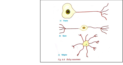
DIAGRAM OF RELAY NEURONE

TYPES OF RELAY NEURONES
i. A unipolar neurone: is a type of neurone that has its axon extending from its cell body.
ii. A bipolar neurone: is a type of neurone that has an axon and dendrons extending in two different directions from the cell body.
iii. A multi-polar neurone: has one axon and several dendrons extending from the cell body in different directions.

NB: The axon extends to the motor neuron
3. MOTOR NEURONES
Are nerve cells that transmit impulses from the central nervous system to the effectors
The cell body of a motor neurone is at one end of the neurone and lies entirely within the central nervous system.
It has tiny branches at each end (dendrites) and a long fibre (axon) that carries the signals or nervous impulses.
Motor neurones are also called efferent neurones
Function of motor neurones
i. To transmit impulses from the central nervous system to the effectors.

STRUCTURAL DIFFERENCES BETWEEN MOTOR AND SENSORY NEURONES
MOTOR NEURONE | SENSORY NEURONE |
| (i) The cell body is located at one end of the neurone | The cell body located near one end of the neurone |
| (ii) It is multipolar | It is unipolar |
| (iii)Has a large and irregular cell body | Has small and definite cell body |
| (iv) Has dendrites surrounding cell body | Has no dendrites which surround cell body |
NERVOUS SYSTEM
This system is made up of the brain, spinal cord and nerves.
Parts of the nervous system
Nervous system is divided into two parts, namely:
1. Central nervous system
2. Peripheral nervous system
1. CENTRAL NERVOUS SYSTEM (CNS)
Is the part of the nervous system consisting of the brain and spinal cord.
It coordinates all the neural functions.
THE COMPONENTS OF THE CENTRAL NERVOUS SYSTEM AND THEIR FUNCTIONS
The central nervous system has two main components, namely:
i. The brain
ii. Spinal cord
THE BRAIN
Is a delicate organ enclosed within a body structure called the skull or cranium.
Brain is the master control of the body.
Brain is covered by a system of membrane called meninges.
The brain is the main centre for integrating and coordinating impulses.
Function of human brain
i. The human brain is a specialized organ that is ultimately responsible for all thought and movement that the body produces.
ii. It allows humans to successfully interact with their environment, by communicating with others and interacting with inanimate objects near their surroundings. For example If the brain is not functioning properly, the ability to move, generate accurate sensory information or speak and understand language can be damaged as well.
PARTS OF THE HUMAN BRAIN
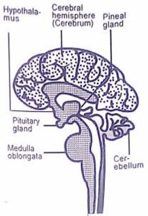
The human brain is divided into three parts, namely:
1. Fore brain
2. Mid brain
3. Hind brain
DIAGRAM OF HUMAN BRAIN

1. FORE BRAIN
Is the anterior portion of the brain
The outer portion is grey hence called grey matter and inner portion is whitish hence called white matter.
Fore brain is responsible for voluntary actions
Fore brain is made up of:
i. Cerebrum.
ii. Hypothalamus
iii. Thalamus
iv. Pituitary gland
v. Olfactory lobes
I. CEREBRUM
Is the largest part of the human brain
Cerebrum is covered by a thin layer of grey matter called cerebral cortex
Functions of the cerebrum
The cerebrum has the following functions:
i. It is responsible for reasoning and intelligence.
ii. It is involved in learning, imagination and creativity.
iii. It is the memory centre.
iv. It is responsible for personality or character.
v. It controls voluntary body movement such as walking and dancing.
vi. It is responsible for sight, hearing, taste, smell and speech.
Parts of the cerebrum
Cerebrum is divided into two parts (cerebral hemispheres), namely:
i. Right hemisphere
ii. Left hemisphere
Right hemisphere
Is the part of the cerebrum which sends and receives impulses from the left side of the body
Left hemisphere
Is the part of the cerebrum which sends and receives impulses from the right side of the body
HYPOTHALAMUS
This part is concerned with body temperature and osmoregulation.
> It contains osmoreceptors and thermoreceptors that detect changes in osmotic pressure and internal body temperature respectively.
> It has a very rich blood supply
Function of the hypothalamus
i. It coordinates and controls the autonomic nervous system
ii. It has centers that control appetite, thirst and sleep.
iii. It also controls the activities of pituitary gland.
iv. It acts as an endocrine gland.
PITUITARY GLAND
This is the master of endocrine glands.
Function of pituitary gland
i. It secretes hormones which control osmoregulation, growth, metabolism and sexual development.
OLFACTORY LOBES
Is the part of fore brain that receives impulses of smell via olfactory nerves from the nose.
Function of olfactory lobes
i. It is concerned with the sense of smell.
2. MID BRAIN
Is the smallest part of the brain which found between the fore brain and hind brain.
The mid brain consists of the optic lobes, which are the main area for audio and visual processing.
Functions of the midbrain
i. To relay information between the fore brain and hind brain.
ii. To relay information between fore brain and the eye through optic nerves.
iii. It is responsible for the movement of the head and trunk.
THALAMUS
The thalamus is located in the middle part of the brain.
i. It helps to control the attention span, sensing pain.
ii. It monitors input that moves in and out of the brain to keep track of the sensations the body is feeling.
iii. It contains the centre for the integration of sensory information.
3. HIND BRAIN
It is made up of cerebellum and the medulla oblongata
CEREBELLUM
Is located in front of medulla oblongata
The cerebellum controls essential body functions such as balance, posture and coordination, allowing humans to move properly and maintain their posture.
Functions of the cerebellum
i. It maintains posture, movement and balance
ii. It ensures that all muscles work together to produce smooth coordinated voluntary movement.
iii. It assists in the learning of new motor skills like playing the piano, swimming and riding a bicycle.
NB: Damage to the cerebellum results in uncoordinated movements
MEDULLA OBLONGATA
This is the central part of the autonomic nervous system
Function of medulla oblongata
i. It controls all unconscious activities of the body e.g. Breathing, heartbeat, digestion, dilation and contraction of blood vessels, secretion of juices from glands and temperature regulation
ii. It contains a number of reflex centre for regulating heartbeat, breathing, blood pressure.
iii. It controls swallowing, salivation, vomiting, coughing, and sneezing.
TOPIC 3: COORDINATION - BIOLOGY NOTES FORM THREE

